You are currently viewing only those items made visible to the public. Click here to sign in and view the full catalogue.

UCL IQPath
| MANUFACTURER | |
|---|---|
| ACRONYM | UCL IQPath |
| RESTRICTIONS | No restriction |
|---|
| CONTACT 1 | Sebastian Brandner |
|---|---|
| CONTACT 2 | ucl.iqpath@ucl.ac.uk |
| Enquire about this item | |
| SITE | Institute of Neurology |
Description
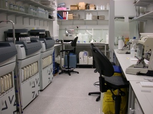
Our state of the art facility performs histological and immunohistochemical techniques on research and diagnostic specimens. Our main expertise is mouse and human histology of the central nervous system, but we cut up, embed, section and stain any other tissue.
We are specialised on paraffin sectioning and all our antibodies are optimised for formalin fixed, paraffin embedded material. If you are interested in frozen sectioning, please enquire at ucl.iqpath@ucl.ac.uk.
Our team can process large quantities (sectioning of up to 100 paraffin blocks per day) and we have the equipment to carry out 150 immunostainings per day. We are offering a large repertoire of antibodies for paraffin sections, (more than 100 antibodies optimised for human and more than 50 antibodies optimised for murine targets). We can also digitise slides, store the data and make them accessible via web browser (see our Digital Pathology section) .
In addition to histological techniques, we advise on histological analysis, in particular of the brain, and the spinal cord, on specific request. The facility is led by Professor Sebastian Brandner, who has expertise in human neuropathology and mouse phenotyping, with particular focus on developmental abnormalities, neuro-oncology and neurodegeneration.
Specification
The following instruments are operational in IQPath Equipment Description Brand Tissue processor LEICA Cassette printer Thermo Embedding centre LEICA Slide label printer Thermo Slide label printer Thermo Microtome LEICA Microtome LEICA Microtome LEICA Waterbath LEICA Waterbath LEICA Waterbath LEICA Automated Immunostainer Ventana/Roche Automated Immunostainer Ventana/Roche Automated Immunostainer Ventana/Roche Robotic Coverslipper LEICA Slide scanner with autoloader LEICA Slidepath Viewing software LEICA Slidepath Teaching software LEICA Image analysis software Definiens File server for Definiens Dell
Associated Items
-
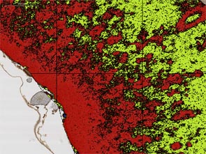
Definiens Tissue Studio software
Image analysis solution for quantitative digital pathology. For tissue quantification by providing morphological fingerprints and biomarker expression profiles on a cell-by-cell basis. Analyses full digital slides Please be advised that this softwar
more details » -
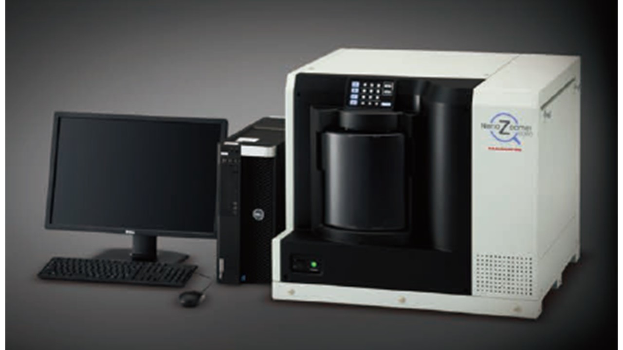
Hamamatsu high throughput slide scanner
The NanoZoomer series is a family of whole slide scanners that rapidly scan glass slides to convert them to digital data. The NanoZoomer S360 is the latest high-throughput model, ideal for uses in hospitals and clinical laboratories. The scanning c
more details » -
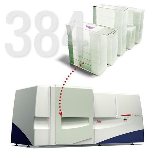
Leica Slidescanner
For digitising histological slides. Please note that this device is approaching end of life. We recommend using our Hamamatsu NanoZoomer for new projects. https://www.research-equipment.ucl.ac.uk/myprofile/items/3827/hamamatsu-high-throughput-slide-
more details » -
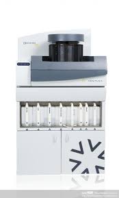
Ventana Immunostainer
Immunostainer
more details »
Item ID #.
Last Updated: 20th January, 2014
There are no publically available categories listed at present. You may have to sign in to browse this catalogue.
